Molecular and General Genetics 256: 661-673
(1997)
Transcripts at the mating type locus of Cochliobolus heterostrophus
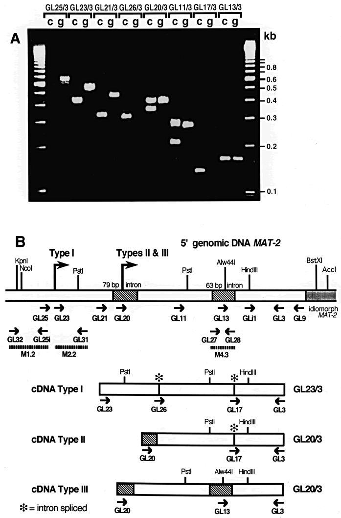 |
Fig. 6A, B Di?erential splicing of introns in the 5¢ leader of MAT-2 transcripts. A Mapping of MAT-2 transcription start sites and di?fferential splicing of the 5'-flank introns. g, genomic DNA; c, cDNA; a 100-bp DNA marker ladder was loaded in each of the outer lanes (fragment sizes indicated on the right). Numbers above the lanes designate primer pairs used (see Figs. 5 and 6B; Table 2). Fragments amplified by PCR from a cDNA template using the 3' primer GL3 and a series of 5' primers (GL25, GL23, GL21, GL26, GL20, GL11, GL17, GL13) are compared to the corresponding products amplified from genomic DNA (Table 2). Note that: (i) the most 5' primer which resulted in MAT-2-cDNA amplification is GL23 and no amplification occurred with GL25; (ii) no fragment was amplified from genomic DNA with 5' primer GL26 or GL17, which are speci®c for the sequence generated after splicing of the 79-bp or the 63-bp intron, respectively (iii) due to di?erential splicing of the 63-bp intron, two fragments were amplified from cDNA with 5' primers located between the 79- and 63-bp introns (GL11) or inside the 79-bp intron sequence (GL20) and (iv) single cDNA fragments were obtained with 5' primers GL23, GL21, and GL26, indicating that when the 79-bp intron is spliced out, the 63-bp intron is also removed. B Classification of MAT cDNAs according to the pattern of splicing of the 79- and 63-bp introns in the 5'-flank of the idiomorph. Maps representing the three cDNA types are compared with genomic 5'-flanking DNA of MAT-2. Type I, Type II and Type III transcription start sites are indicated above the genomic DNA (large arrows). Locations of PCR primers (small arrows), non-spliced introns (hatched boxes), spliced introns (asterisks) and PCR fragments (plus primers used to generate them) used as templates for RNA probes (thin broken bars) are indicated. The three cDNA Types are represented by cDNAs GL23/3 and GL20/3 |
| Article in PDF format (540 KB) | Abstract Fig. 1 Fig. 2 Fig. 3 Fig. 4 Introns Fig. 5 Fig. 6 Fig. 7 Fig. 8 |
|
|
The Seed Biology Place
|
Webdesign Gerhard Leubner 2000
|