Darwin Review - Journal of Experimental Botany 62: 3289-3309 (2011)
First off the mark: early seed germination
Department of Biological Sciences, Simon Fraser University, 8888, University Drive, Burnaby BC, V5A 1S6, Canada (K.M.)
*Joint first authors: K.W., K.M.
Received December , 2010; accepted February 23, 2010; published online April 12, 2010
DOI 10.1111/j.1469-8137.2010.03249.x

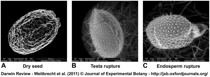
Figure 4. Scanning electron microscopy (SEM) and environmental scanning electron microscopy (eSEM) images of Arabidopsis thaliana seed germination.
(A) Air-dry seed of Arabidopsis (SEM) showing the hexagonal testa cells on the surface with the mucilage packed into the middle elevation resulting in the columella.
(B) Imbibed Arabidopsis seed (eSEM) in testa rupture state; the micropylar endosperm covering the radicle is visible.
(C) Arabidopsis seed (eSEM) in endosperm rupture state; the emerged radicle is visible and designates the completion of germination. eSEM works without freezing, coating, fixing or embedding and in a relative humidity of ≥ 90 %. It can thus be used to image a living organism with high magnification (Muscariello et al., 2005; Windsor et al., 2000).
Images taken by Dr. Ralf Thomann, Freiburg Materials Research Center (FMF).
| Article in PDF format (900 KB) |
|
|
|
The Seed Biology Place |
Webdesign Gerhard Leubner 2000 |